
Congress Schedule
A group of sessions will be released at 8:00am and 4:00pm in Japan Standard Time(JST) from October 4 to 6.
Please check what time your local time will be.
As of August 18, 2020
It is subject to change.
| Time | Track #1 |
Track #2 |
Track #3 |
Track #4 |
Track #5 |
Track #6 |
Track #7 |
||||||||||||||||||||||||||||||||||||||||||||||||||||||||||||||||||||||||||||||||||||||||||||||||||||||||||||||||||||||||||||||||||||||||||||||||||||||||||||||||||||||||||||||||||||||||||||||||||||||||||||||||||||||||||||||||||||||||||||||||||||||||||||||||||||||||||||||||||||||||||||||||||||||||||||||||
|---|---|---|---|---|---|---|---|---|---|---|---|---|---|---|---|---|---|---|---|---|---|---|---|---|---|---|---|---|---|---|---|---|---|---|---|---|---|---|---|---|---|---|---|---|---|---|---|---|---|---|---|---|---|---|---|---|---|---|---|---|---|---|---|---|---|---|---|---|---|---|---|---|---|---|---|---|---|---|---|---|---|---|---|---|---|---|---|---|---|---|---|---|---|---|---|---|---|---|---|---|---|---|---|---|---|---|---|---|---|---|---|---|---|---|---|---|---|---|---|---|---|---|---|---|---|---|---|---|---|---|---|---|---|---|---|---|---|---|---|---|---|---|---|---|---|---|---|---|---|---|---|---|---|---|---|---|---|---|---|---|---|---|---|---|---|---|---|---|---|---|---|---|---|---|---|---|---|---|---|---|---|---|---|---|---|---|---|---|---|---|---|---|---|---|---|---|---|---|---|---|---|---|---|---|---|---|---|---|---|---|---|---|---|---|---|---|---|---|---|---|---|---|---|---|---|---|---|---|---|---|---|---|---|---|---|---|---|---|---|---|---|---|---|---|---|---|---|---|---|---|---|---|---|---|---|---|---|---|---|---|---|---|---|---|---|---|---|---|---|---|---|---|---|---|---|---|---|---|---|---|---|---|---|---|---|---|---|---|---|---|---|---|---|---|---|---|---|---|---|---|---|---|---|---|---|---|---|---|---|---|---|
| All sessions in this time slot will be released at 8 am JST. |
Accelerated Tooth MovementAccelerated Tooth Movement
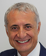
Dr Darendeliler is Professor and Chair of Orthodontic, Discipline of Orthodontics, at the University of Sydney and Head of Department, Orthodontics, Sydney Dental Hospital, Sydney South West Area Health Service. He received his dentistry training from the University of Istanbul and his PhD from the University of Gazi, in Turkey and his first specialist training in orthodontics from the University of Geneva, Switzerland and his second specialist training from the High Education Council, Turkey. During the course of his career he has undertaken duties as a clinical instructor, research and postgraduate coordinator (Maitre d'Assisstant et de Recherche) at the University of Geneva, Assistant Professor at the University of North Carolina, Research Professor at the University of Southern California. Dr Darendeliler has been recognized for his efforts with multiple prizes including the Sheldon Friel Award, the highest recognition from the European Orthodontic Society, the Begg Award, the highest research award from the Australian Society of Orthodontists, and the Huston award for best research from the European Orthodontic Society. His research interests include orthodontic tooth movement, root resorption, obstructive sleep apnoea, temporary anchorage devices, sequential aligners, self-ligating brackets, orthopaedic treatment modalities, accelerated tooth movement, magnetic fields and forces and dentofacial orthopedics. He lectured in North and South America, Europe, Asia, Africa and Australia. In addition to his research and teaching commitments he also maintains a private specialist orthodontic practice. Abstract Clinicians have been looking at the ways of accelerating tooth movement for several reasons: Patient's demand due to increase in number of adult patients, to avoid problems related to treatment duration such as demineralization, periodontal issues and root resorption, to increase the productivity, profitability and efficiency, to have better anchorage control and to differentiate themselves from other practitioners. Several methods were used to accelerate tooth movement however the benefits of the methods used have to be looked at in terms of side effects and clinical significance. The only way to accelerate the orthodontic tooth movement is to accelerate bone resorption via biological response from the periodontal ligament as a result of mechanical loading. In fact the question to be asked is: Accelerated bone resorption; What is the evidence? Several methods, drugs and external stimuli were tested over the years and most of these were promoted by companies usually with little or no evidence. The lecture will address all available methods used to accelerate orthodontic tooth movement and will discuss their validity based on evidence. Accelerated Tooth Movement
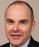
Martyn Cobourne is Professor and Head of Orthodontics at Kings College London and Honorary Consultant in Orthodontics at Guys and St Thomas NHS Foundation Trust. His research is primarily focused on investigating the role of molecular signaling pathways during early craniofacial development. However, he is also interested in the effectiveness of contemporary orthodontic treatment and has led a number of multi-centre clinical trials investigating treatment efficiency. His clinical research won the B. F. and Helen E. Dewel Award of the American Association of Orthodontists in 2019. He has published over 145 peer-reviewed articles and is the author of two successful orthodontic textbooks. He was Director of Research at the British Orthodontic Society between 2012 and 2016 and is currently Editor in Chief of the Journal of Orthodontics. He is an elected member of the Board of Faculty of Dental Surgery at the Royal College of Surgeons of England and a full member of the North Atlantic Division of the USA Angle Society. Abstract A key goal for all orthodontists is the minimisation of treatment duration whilst maintaining excellence of treatment outcome. In recent years, a number of techniques have been popularised that aim to reduce orthodontic treatment time by speeding up rates of tooth movement. These include the use of different bracket designs and archwire sequences, customised appliances, vibrational force application, photobiomodulation and surgical-assisted orthodontics. This lecture will review the scientific basis of these different techniques and focus on the clinical evidence base for their efficiency. The relevance of these findings will be discussed in relation to current clinical practice. |
AirwayAirway
Professional Experience and Education
Honors and Awards
Research Interests Mid-facial expansion, 3D image analysis, Genomewide Association Study of Craniofacial Phenotypes, Finite Element Model (FEM), Applications of 3D Printing, Orthopedic Correction, Airway Changes, Accelerated Tooth Movement, and Micro-implant (MI) Design study Abstract The primary aim of this presentation is to illustrate how the maxillary skeletal expander (MSE) has evolved from its birth, by examining the dental and skeletal effects of maxillary expansion when the conventional rapid palatal expander (RPE), the surgically-assisted rapid palatal expander (SARPE), and the micro-implant assisted MSE are used. Dental expansion, bone bending, and true skeletal expansion will be compared. The adverse clinical consequences of RPE and SARPE will be explored, and a new approach eliminating these problems by the use of MSE will be presented. Clinical cases involving non-surgical midfacial expansion in both adolescent and adult patients will be examined. Airway
Professional Experience and Education
Honors and Awards
Research Interests
Abstract Orthognathic surgical procedures are a reliable method of correcting dentoskeletal deformities of the jaws. These procedures have shown good long term stability in two dimensions. However, three-dimensional assessment of the long term stability of bimaxillary surgery is lacking in the literature. Airway
Professional Experience and Education Dr. LIU Yuehua is a Professor and Dean of Shanghai Stomatological Hospital, Fudan University. He got the Ph. D degree in 1996 from Peking University and was a Postdoctoral fellow in the University of British Columbia, Canada from 1998 to 2000. Honors and Awards Now he is an Executive Member of the Council and Vice-President of Chinese Orthodontic Society of Chinese Stomatological Association (CSA), Vice-Director of the Council and Immediately Past President of Shanghai Orthodontic Society of Shanghai Stomatological Association (SSA), an Examiner in Orthodontics, the Royal College of Surgeons of Edinburgh.Professor LIU has been in charge of 6 General Programs of National Natural Science Foundation of China (NSFC), published over 30 peer-reviewed papers on SCI Journals and authorized 4 National Invention patents. In addition, he was awarded the second prize of the advanced science and technology, Ministry of Education, PRC, in 2002 and the second prize of the advanced medical science and technology, Chinese Association of Medicine, PRC, in 2003. Research Interests He has been interested in rensearch in obstructive sleep apnea in children, dentofacial vertical control for profile improvement, and lingual orthodontics. Abstract The etiologic factors of Obstructive sleep apnea and hypopnea syndrome(OSAHS) include anatomic narrowing and dysfunction of uppe airway. The pathogenesis,treatment stratege and prognosis of OSAHS in children could be different from that in adult. The manifestation of those children with OSAHS may include hypertrophy of tonsils and adenoid, mouth breathing, narrowed upper arch, clockwise rotated mandilbe,and uppe airway narrowing. Tonsillectomy and adenoidectomy could never eliminate the symptoms of OSAHS in children because hard tissue anatomic abnormality surrounding uppe airway still exist. In this presentation, the children with different sererity of OSAHS were subdivided into several groups with different therapies. those therapeutic methods include Tonsillectomy & adenoidectomy, oral appliance for upper arch expansion and mandibular advancement, and myofunctional reeducation. the subjective and objective treatment efficency to OSAHS were assessed by using questionnaire, and polysomnography. The effects of dentofacial growth modification from oral appliances and myofunctional reeducation were measured and analysed. The results demonstrated that combination of various methods may achievevd more satisfied curative effect. |
Patient's Expectations and TreatmentPatient’s Expectations and Treatment
Professional Experience and Education
Honors and Awards
Research Interests Orthodontic Clinical Outcomes Abstract The specialty of orthodontics is undergoing rapid transformation and the pace of change has left many orthodontists feeling uncertain and perhaps even afraid of what the future will hold. While scientific research and appliance innovations allow achievement of increasingly dramatic and precise dental movements, patients often request faster and more convenient treatment options that lead to compromised outcomes. Technological advancement, economic pressure, increased competition, and shifts in marketing make it difficult to predict where we are going. It is clearly established that most patients seek orthodontic treatment because they want their teeth and their smile to look better. Is it appropriate to set goals for orthodontic treatment if there are no confirmed detrimental effects caused by not meeting them? What do our patients expect from the orthodontic treatment they voluntarily seek, and how willing are they to accept compromises? The answers to these questions may shape the future of orthodontics as we adapt to a changing environment. Patient’s Expectations and Treatment
Professional experience and education
Honors and Awards
Research interests Outcomes of orthodontic and orthognathic treatment Abstract Understanding and managing patient expectations are fundamental aspects of any healthcare intervention, but are particularly important in elective treatments such as orthodontics. |
Imaging and Digital OrthodonticsImaging and Digital Orthodontics
Chief Centre for Dental Education and Research All India Institute of Medical Sciences New Delhi from 2015 Orations Indian Orthodontic Society Oration 54th Indian Orthodontic Conference Bhubaneshwar 2019 Honours and Awards Shakuntala Amirchand Award by the Indian Council of Medical Research. 1994 Research interests 3D Technology in Orthodontics NonExtraction Treatment Miniscrew Implants Cleft lip and palate Abstract The science and technology of 3D imaging at relatively low radiation and advanced software capabilities have enabled clinicians and researchers three dimensional visualisation, measurements and volumetric analysis of the craniofacial region. Researchers have proposed new 3D landmarks and 3D cephalometric analysis. The complexity of on-screen three-dimensional landmark plotting requires considerable effort and time notwithstanding the experience of the operator as compared to landmark plotting on 2D conventional cephalogram. Such issues necessitated the urge for automating the process of 3D landmark plotting and measurements. In the last few years, three types of approaches for automatic detection of cephalometric landmarks have been proposed. The first being registration-based and second knowledge-based and third deep learning-based. We have developed an innovative knowledge-based technique and tested its reliability for automation of useful 3D cephalometric landmarks. The automated detection can also generate linear, angles and ratios measurements. One step ahead, using template matching extended to a knowledge of anatomical definitions on CBCT, our group has developed an automated volumetric analysis of paranasal sinus and nasal-respiratory passages. Imaging and Digital Orthodontics
Prof Nikhilesh R. Vaid is currently the PRESIDENT -ELECT of the World Federation of Orthodontists. He is a Past President of the Asian Pacific Orthodontic Society & the Indian Orthodontic Society. Prof Vaid is Editor in Chief of APOS Trends in Orthodontics -the Journal of the Asian Pacific Orthodontic Society and three issues of Seminars in Orthodontics, including one on "Digital Technologies in Orthodontics" Abstract The past decade in Orthodontics has made some very "emphatic statements",which will chart the course of how orthodontics is practiced, marketed, researched and taught over the next decade and probably the rest of this century! Imaging and Digital Orthodontics
Professional Experience and Education
Honors and Awards
Research Interests Dr. Toru Deguchi has been focusing his clinical interests in the field of temporary anchorage device, lingual orthodontics, and interdisciplinary cases. He has been also engaged in the research field such as histomorphometric studies related to tooth movement and pain related studies. Recently, he has initiated research related to three-dimensional diagnostic system in clinical orthodontics. Abstract In recent years, there have been a significant progress in three-dimensional (3D) diagnostic system in the field of orthodontics. Remarkable development of intra-oral scanner, the use of digital model, 3D printer, and cone beam CT has contributed to these innovative changes. Combining these 3D tools will be able to visualize the problem and help patients understanding the treatment goals and may contribute in enhancing the work efficiency for the orthodontists. These techniques also have influenced in constructing various orthodontic appliances including in-house aligners and retainers. In this presentation, I would like to introduce how we apply these 3D systems in diagnosis and treatment planning such as setup models, visual treatment objectives, and managing surgical prediction and constructing the splints in our post graduate orthodontic program. I will also present some of our current research with regard to reliability and the usefulness of 3D diagnostic system in clinical orthodontics. |
Cleft Lip and PalateCleft Lip and Palate
University of Helsink i and Helsink i University Hospital. In this talk , I will present some of the dramatic advances that have shaped our understanding of how the facial processes develop and our current k nowledge of the causes of cleft lip and palate. I will describe the normal development of the craniofacial region including the lips and palate emphasising the critical stages and the k ey regulatory mechanisms that can go wrong and result in a craniofacial deformity. Cleft Lip and Palate
Professional Experience and Education
Honors and Awards
Research interests My research interests are centred around children born with orofacial clefting. I supported the National Institute for Health Research funded programme, Cleft Care UK, where we evaluated the impact of the centralisation of cleft services following the recommendations made by the Clinical Standards Advisory Group CSAG in 1998. I lead the Scar Free Foundation birth cohort gene backed study known as the Cleft Collective. Abstract The care of children born with a cleft lip and palate requires multidisciplinary care from a wide range of healthcare professionals and arguably, as this is a repairable structural anomaly, the surgeons (from whatever parent discipline) would be expected to be pivotal in organising healthcare of these children. Cleft Lip and Palate
Abstract Cleft lip and palate (CLP) causes various morphological and functional problems in esthetics of nose and lip, feeding, speech and occlusion. Among them, correction of occlusion requires long and comprehensive intervention. It is important to minimize the burden of orthodontic treatment, together with providing successful outcome for patients with CLP. The condition of intermaxillary relation, maxillary arch form, and normal tooth eruption are all related to the difficulty of orthodontic correction. A tight collaboration of orthodontists with other specialists in related fields is essential to reduce the burden of CLP children. |
Genetics
Professional Experience and Education
Honors and Awards
Research Interests My current efforts also broadly focus on gene discovery and phenotype dissection of dentofacial variation and eruption disorders using 2 and 3 dimensional methods for rigorous clinical characterization, genotyping and mutational analysis through the candidate gene approach. Abstract This lecture will provide an overview of current practices in the diagnosis and treatment of malocclusions with a genetic basis. We are quickly approaching a time when personalized orthodontics will be an integral part of our diagnostic regime just as it is with medicine. Taking a family history in fact represents the gold standard in the diagnosis and management of medical (and by extension) dental disorders. The objective of this lecture will be to recognize and diagnose both common and rare dental disorders encountered in orthodontic practices from both clinical and genetic perspectives. Data from genetic and clinical studies have helped to create a paradigm shift in contemporary orthodontic practices. Applying genetic knowledge to the field of orthodontics will augment the current differential diagnosis of dental disorders, permitting recognition of etiologically distinct disorders that respond to treatment in different ways. That is, proper diagnosis equals proper treatment. This session will include discussion of Primary Failure of Eruption, ankylosis, delayed eruption and Class III malocclusion with an emphasis on adopting a diagnostic rubric for proper management. Or, what to do, and what not to do. Genetics
CV Prof. dr. Carine CARELS, DDS, PhD, hon fellow FDSRCS
Abstract Tooth agenesis and orofacial clefts both represent very common developmental anomalies and their co-occurrence is often reported in patients as well as in animal models. Here we aimed to perform a systematic review to thoroughly investigate the literature in order to identify genes and genomic loci contributing to syndromic or non-syndromic co-occurrence of tooth agenesis and orofacial clefts and to gain insight into the molecular mechanisms underlying their dual involvement in the development of teeth and facial primordia. Altogether, 84 articles including phenotype and genotype description provided 9 genomic loci and 26 gene candidates underlying the co-occurrence of the two congenital defects: MSX1, PAX9, IRF6, TP63, KMT2D, KDM6A, SATB2, TBX22, TGFα, TGFβ3, TGFβR1, TGFβR2, FGF8, FGFR1, KISS1R, WNT3, WNT5A, CDH1, CHD7, AXIN2, TWIST1, BCOR, OFD1, PTCH1, PITX2 and PVRL1. Genetics
Professional Experience and Education
Honors and Awards
Research Interests Human genetics in orofacial disease and traits Abstract Malocclusion, an incorrect relationship between the maxilla and the mandible or a general misalignment of the teeth, is highly influenced by genetics. Genetic exploration of human craniofacial morphology, as morphological variation, began in 2001. In recent years, several genome-wide studies have surfaced that began with simple curiosity regarding the development and organization of the human face. Major contributions to progress in this area were made by researchers in various fields, including anatomy and human evolution, rather than by dentists. Explosive recent advances in genome science have identified genetic factors of malocclusion in humans. Research on the genetics of human mandibular prognathism was initiated in 2005; thereafter, it progressed by studying human genes with malocclusion as a phenotype. |
APOS Lectures
Professional Experience and Education
Honors and Awards
Research Interests Adult orthodontics Abstract How Invisalign change the orthodontics? As we know, Invisalign was first released by Align Technology in 1997. After 23 years' growth and development, the Invisalign has changed the life of our orthodontists. The Invisalign has changed the way how the orthodontists treat the patients, and also the way how the orthodontists work at the clinic. At the same time, The Invisalign has changed the way how the patients come to see the orthodontists. The Invisalign has made the orthodontic treatment more invisible and more comfortable. The Invisalign has also made the opportunity for those patients who couldn't receive the orthodontic treatment with conventional braces.
Professional Experience and Education
Abstract As intraoral scanners and digital workflows gain popularity in clinics everywhere, clinicians are able to increase their clinic offering with in-house clear aligners and differentiate themselves from the rest.
Professional Experience and Education
Honors and Awards
Research Interests Biology of periodontal tissues Abstract We investigated an easy debonding method for ceramic brackets using a light-cured Bis-GMA resin adhesives containing thermal expansion microcapsules and CO2 laser. |
||||||||||||||||||||||||||||||||||||||||||||||||||||||||||||||||||||||||||||||||||||||||||||||||||||||||||||||||||||||||||||||||||||||||||||||||||||||||||||||||||||||||||||||||||||||||||||||||||||||||||||||||||||||||||||||||||||||||||||||||||||||||||||||||||||||||||||||||||||||||||||||||||||||||||||||||
Evidence Based OrthodonticsEvidence Based Orthodontics
H-Index 53 Google Scholar - 32 Scopus - 31 Web of Science Education DDS (Universidad Peruana Cayetano Heredia ? Peru - 1994) Employment Tenured Professor at the University of Alberta Extramural Private Practice in Edmonton, Canada Current Position Orthodontic Program Director, University of Alberta (since 04/10) Presentations More than 120 international presentations around the topics of Clinical Orthodontics, Evidence-based Dentistry and Evidence-based Orthodontics (Argentina, Australia, Bolivia, Belgium, Brazil, Canada, Colombia, Chile, Costa Rica, Germany, Iceland, Israel, Italy, Mexico, Netherlands, Panama, Paraguay, Peru, Poland, Romania, Uruguay, USA, Scotland, Switzerland and Spain) Major Teaching awards 2014 Department of Dentistry, Gibb Teaching Scholar Award, 2010 Faculty of Medicine and Dentistry, University of Alberta ? Tier II Clinical Award for Excellence in Mentoring Student, 2007 Association of Canadian Faculties of Dentistry (ACFD) - W. W. Wood Award for Excellence in Dental Education Publications 6 book chapters related to Evidence-based Dentistry 280 peer-reviewed articles and 37 commentaries published. Abstract This presentation will explore how recently published evidence (systematic reviews and randomized clinical trials) focused on impacted teeth has impacted my day to day clinical decisions when faced with a potential impacted canine. The following questions will be explored: Evidence Based Orthodontics
Professional Experience
Honours and Awards
Research Interests Treatment effectiveness and Predictability Abstract While there is increasing acceptance of the necessity for indefinite retention, there has been relatively little prospective research concerning the relative effectiveness of fixed and removable retention protocols in the longer term. |
Obstructive Sleep ApneaObstructive Sleep Apnea
Dr. Juan Martin Palomo is a professor and the Orthodontic Residency Director at Case Western Reserve University in Cleveland, Ohio. Dr. Palomo's contributions to craniofacial imaging, informatics, and airway analysis have been recognized through medical and dental research awards, national and international presentations, and numerous peer reviewed publications. At the American Association of Orthodontists (AAO) he is a member of the Council on Scientific Affairs, member of the AAOF Planning and Awards Review Committee, the AAO representative for the Imaging Gently Healthcare group, and past chair of the Committee on Technology. He is also the Associate Editor for the Innovations and Technology section of the AJODO. He was a member of the AAO task force that resulted in a white paper on sleep apnea and orthodontics. Dr. Palomo is a board certified orthodontist, and Angle Society member, and an active member of the American Association of Orthodontists, the American Academy of Oral and Maxillofacial Radiology, and the American Academy of Sleep Medicine. Abstract Traditionally we are trained to see and analyze the airway from only the lateral view on a cephalometric film. But the airway is a three-dimensional (3D) structure, and that third dimension may be hiding something relevant to our diagnosis. This presentation will highlight some of the airway research from Case Western Reserve University, and will show how different commercially available programs can be used to analyze the airway volumetrically, how the airway is different for different craniofacial situations, and what happens to the airway when we extract or expand as part of our treatment plan. With more than 80 respiratory disorders, the orthodontist can play a big role helping children and adults, specially regarding Obstructive Sleep Apnea (OSA). This presentation will show how the orthodontist can help identify and manage OSA in pediatric and adult patients, from using oral appliances, to surgical movement of the jaws, and newest hypoglossal stimulation methods. When moving from 2D to 3D, distances and angles turn into areas and volumes, and understanding the airway may take orthodontics to the next level, increasing the scope of what can be done clinically. Obstructive Sleep Apnea
DDS, MSD, PhD degrees from Seoul National University (SNU) Abstract Obstructive sleep apnea (OSA) is a sleep disorder due to repetitive episodes of upper airway collapse. Several treatment modalities have been used for OSA patients including diet, exercise, sleep position change, nCPAP, nasopharyngeal soft tissue procedures, genioglossus advancement, oral appliances, rapid palatal expansion appliances, maxillomandibular advancement (MMA), hypoglossal nerve stimulation, and tracheostomy. |
Diagnosis and Treatment PlanningDiagnosis and Treatment Planning
Dr Hong He, BDS, MDS, PhD Abstract Pediatric sleep-related disorder breathing (SDB) is caused by upper airway obstruction, resulted often from adenoid hypertrophy, tonsil hypertrophy or nasal disease. Mouth breathing is one of the most common clinical manifestations of pediatric SDB. Children whose mouth breathing remains untreated may develop long, narrow faces, high palatal vaults, dental malocclusion, gummy smiles etc. because of orofacial muscle imbalance. SDB in children has been associated with a variety of comorbidities, including failure to thrive, hypertension, attention deficit disorder, enuresis etc. Orthodontists who have the knowledge of SDB can play an important role in the early diagnosis and treatment of pediatric SDB. It is recommended that the orthodontist perform a clinical risk assessment for SDB. Orthodontists may be involved in the treatment of pediatric SDB by correcting the underlying dental and skeletal deformities. When making the orthodontic treatment plan, orthodontists should account for children's upper airway condition. Diagnosis and Treatment Planning
Career History
Award
Writing
Abstract This presentation consists of four parts. |
TMJ
Professional Experience and Education
Honors and Awards
Research Interests Temporomandibular disorders Abstract Temporomandibular disorders (TMDs) are frequently accompanied with malocclusion in orthodontic patients. Severe mandibular condylar resorption causes retrusion and deviation of the mandible, leading to the onset and progress of malocclusions such as skeletal open bite. In our department, if TMDs are diagnosed during the initial clinical examination of malocclusion patients, Temporomandibular joint (TMJ)-MRI is taken as a general rule. In cases where mandibular condylar deformity is suspected, orthodontic treatment will not start unless the condylar resorption abates. TMJ
Professional Experience and Education
Honors and Awards
Research Interests Hormonal Pathobiology of TMJ Degeneration Abstract The pre-treatment or in-treatment identification of patients undergoing idiopathic condylar resorption of the TMJ and their management remains one of the biggest challenges in orthodontics. Frequently, because these joint resorptive events and associated adverse dental and skeletal alterations occur during orthodontic treatment, the concomitant compromised function and facial changes are often attributed to the orthodontic therapy itself. Much is still unknown about these perplexing conditions. In his presentation, Dr. Kapila will discuss the current understanding of the pathogenesis, diagnosis and management of idiopathic condylar resorption, including that which may occur during juvenile idiopathic arthritis. Early recognition through careful history taking and clinical examination together with newer diagnostic methods using 3D imaging and biomarkers and constant monitoring can help mitigate some of the challenges arising during orthodontic treatment. Alternatively, these findings can be used to defer treatment of these patients until stabilization occurs. Specific radiographic and other features that indicate cessation of the disease process are a prerequisite for initiating orthodontic therapy. Dr. Kapila will also discuss a range for patient-specific treatment options for individuals presenting with different phenotypic and degenerative TMJ findings in idiopathic condylar resorption ranging from conservative management to orthognathic surgery. |
Craniofacial Growth and DisordersCraniofacial Growth
Professional Experience and Education
Research Interests
Abstract Scientific background of orthodontic-orthopedic treatment for skeletal malocclusion during growing period is one of the important issues to be addressed. Mandibular condylar cartilage (MCC) is the major growing cite of mandible by endochondral bone formation. We have been performing series of experiments to analyze the cellular response of differentiating chondrocyte to compressive and tensional stress loading. In the first experiment, electrical stimulation of lateral pterygoid muscle inhibited the chondrogenic differentiation of mesenchymal cells in the posterior area of MCC in growing rats. Since distribution of mechanicals stress in temporomandibular joints was unclear, we employed midpalatal suture cartilage (MSC) in growing rats to verify the hypothesis that compression enhances chondrogenesis and tension inhibits chondrogenic differentiation in the MSC. As the result, hypertrophy of chondrocytes was enhanced in the MSC compressed by orthopedic force, while the cartilage was replaced by bone under expansive force. Further, integrins are found to be expressed in the MSC and Extracellular Signal-regulated Kinase (ERK) was activated by expansive force. To analyze the cellular mechanisms of mechano-response of differentiating chondrocytes, embryonic limb bud cells were mechanically stimulated in vitro. When mouse limb bud cells were compressed in collagen gel 3-D culture, differentiation of chondrocytes was accelerated with enhanced expression of Sox9, the transcriptional activator for cartilage specific collagen, Col2a1. On the other hand, expansive stress inhibited the chondrogenic differentiation of rat limb bud cells in stretched micromass culture through integrin mediated ERK signaling pathway. Thus, different types of mechanical stress would differentially regulate the differentiation of chondrocytes. Craniofacial Growth
Professional Experience and Education
Honors and Awards
Research Interests Biomechanics and behaviors of the human craniomandibular complex with special interests in the TMJ, jaw muscles, and tooth movement. Abstract Mechanical loading is important to the growth and maintenance of the secondary cartilages of the condyle and eminence of the temporomandibular joint (TMJ). The fibrocartilage disc between the condyle and eminence distributes loads, provides lubrication and, because it is avascular, depends on mechanical loading for nutrient exchange. Hence, therapies to modify jaw growth and prevent or ameliorate degenerative joint disease could be improved by understanding the mechanical loading conditions within the TMJ. Studies in humans where the magnitude and frequency of TMJ loading were measured to distinguish dentofacial phenotypes and predict longitudinal TMJ change will be highlighted. Key measurements of mechanical loading conditions are the concentration of work input to articulating tissues during jaw use, known as energy density, and the percentage of time that muscles are used to load the jaws out of a total recording time, known as duty factor. These measurements, combined into a mechanobehavioral score (MBS = (TMJ energy densities)2 X jaw muscle duty factors), are being applied to understand better the mechanisms involved with condylar growth and the susceptibility of the TMJ to degenerative changes. This presentation will describe how clinical treatment records and data collected via validated techniques, including numerical modeling, dynamic stereometry, and laboratory and in-field electromyography are employed to determine mechanobehavioral score. Future clinical approaches that could modify mechanobehavior to achieve more successful and predictable orthopedic therapies in children with jaw discrepancies and measures to prevent degenerative changes in the TMJ will be discussed. Craniofacial Growth
Prof. Dror Aizenbud is a full professor of orthodontics and the chairman of the Department of Orthodontics and Craniofacial Anomalies and the deputy director of the School of Graduate Dentistry, Rambam Health Care Campus and Technion Israel Institute of Technology – Faculty of Medicine Haifa, Israel. He is the past president of the IADR Israeli Division. He honored a visiting professor nomination at the Craniofacial Anomalies Center, Stanford University Hospital, California, USA. Prof Aizenbud holds a Master of Science degree, a degree in dentistry and a certificate in orthodontics, both from the Hebrew University and Hadassah School of Dental Medicine, Jerusalem, Israel. He spent a fellowship program in the Craniofacial Center at the University of Illinois at Chicago with the group of Drs. Figueroa and Polley. Abstract Congenital craniofacial anomalies can range from small isolated areas to extensive areas of bone loss involving the entire jawbone and teeth. The resulted malocclusion caused by severe skeletal and dental discrepancies pose a significant challenge for the orthodontist and other health care providers. Nowadays advanced three dimensional technologies of planning and performance of orthodontics, dentofacial orthopedics, distraction osteogenesis and orthognathic surgery type of managements has become a routine care for the growing child to deliver successful treatment results.
Patients with these conditions often presented with functional and esthetic issues call for coordinated surgical, dental and medical multidisciplinary approach. Currently practice standards of patients with congenital craniofacial defects are achieved in a team setting and usually span the first two decades of life using the major advantage of patient's growth potential. |
Bone BiologyBone Biology
Professional experience and education
Honors and Awards Alice L. Jee Memorial Award for junior investigators. International Sun Valley Workshop on Hard Tissues, Sun Valley, Idaho, USA, 1998 Research interests Biology of tooth movement, biomechanics, craniofacial anomalies Abstract The achievement of a planned tooth movement is the result of the interaction between variables related to the orthodontist and variables related to the patient. The orthodontist plans the individual biomechanical system in relation to the centre of resistance of one tooth or groups of teeth. It is known that the location of the centre of resistance varies according to the patients' anatomical characteristics; the anatomy of the supporting tissues and the shape of the roots. The quality of the supporting tissues in terms of bone density has seldomly been taken into consideration as potential variable that could influence the location of the centre of resistance. Surgically facilitated orthodontic tooth movement is one of the various attempts of orthodontists to influence patients' response to enhance tooth movement rate. The principle is based on the local acceleration of bone remodelling activities occurring in bone repair processes, where bone density is initially decreased. Accelerated bone turnover in animal models has shown to increase tooth movement rate and the application of a single force induces a controlled rather than an uncontrolled tipping, as would under normal bone turnover conditions. A Finite Element analysis of tooth movement after corticotomy revealed that translation in corticotomized teeth occurs at a larger M/F ratio compared to normal bone turnover. In order to achieve this, a bigger moment needs to be inserted into the wire. Bone Biology
Professional Experience and Education
Honors and Awards
Research Interests Biological analysis of tooth movement Abstract In a variety of scientific fields, it is a worthwhile topic to visualize natural phenomenon. Newly developed visualizing method often leads breakthrough in the scientific fields. Especially, in the biological field, it is significant to reveal temporal-spatial response happened in the cells with visualizing molecular level phenomenon. Such visualization could provide information to understand cellular behavior to their extracellular stimulus in vivo and in vitro. Although osteocytes are the most abundant cells in bone, it has been difficult to study their biological feature because they are embedded in hard bone tissue. So, even the real 3D structure of the osteocyte was not uncovered till lately. As it is already known orthodontic tooth movement requires dramatic bone changes. Osteocytes are thought to be primary mechanosensory cells. In this presentation, we will introduce our application of confocal laser scanning microscopy, ultra-high voltage electron microscopy and focused ion beam scanning electric microscopy to reveal the bone microstructure and discuss the mechanosensitivity of the cells in bone. Bone Biology
Dr. Sarandeep S. Huja, DDS, PhD currently serves as the Dean, of the James B. Edwards College of Dental Medicine and Professor, Department of Orthodontics Medical University of South Carolina Dr. Huja initially received his dental (1987) and orthodontic (1992) training from the Government Dental College and Hospital, Bombay. From 1993-2001, he was associated with three US institutions, receiving his MS (1995) from Marquette University, Milwaukee, his PhD (1999) and Orthodontic Certificate (1999) from Indiana University, Indianapolis and his DDS (2001) from University of Nebraska Medical Center College of Dentistry, Lincoln. His PhD was conducted under the mentorship of Dr. David Burr from the Department of Anatomy and Dr. W. Eugene Roberts in Orthodontics from Indiana University. From 2001-2011, he was a faculty at the Ohio State University working under the mentorship of Dr. Katherine Vig, a world renowned orthodontist and educator. In 2011 he moved the University of Kentucky as Division Chief of Orthodontics. In 2016 he was appointed the Program Director of Orthodontics and Associate Dean for Faculty and Student Development and Graduate Studies at the University of Kentucky. Abstract Many methods to expedite orthodontic tooth movement have been developed with little promise for current practice. However, the research has led to an affirmation that the benefits of reduction in treatment time is a worthy goal. We will describe and compare animal models to study tooth movement with the focus on tissue level bone biology and relate this fundamental information to tooth movement in humans and clinical practice. Forces for orthodontic tooth movement remain undefined. We will expand on the need to understand the force and engineering stress to produce optimal tooth movements and thus expediting/optimizing tooth movement. In the future, orthodontic tooth movement are likely to be individualize in a patient based on tooth root surface area, anchorage segments, desirable versus undesirable tooth movements, besides gender/age thereby producing direct path and personalized tooth movements for each patient. The participant will thus be exposed to most recent concepts and evidence based methods to achieving orthodontic tooth movements. |
Allied Health Professional Program: TechniciansAdvancement Digital Technology Technicians: Advancement Digital Technology
Professional Experience and Education
Award
Research Interests Biomaterials Abstract The use of digital technology in orthodontics has been increasing in the last few decades. In particular, digital 3D model generated by intraoral scanning has been widely accepted by the field of clinical orthodontics, and is advantageous due to the compact storage space, simplified method of transferring data, potential to expand application for treatment planning, and fabrication of customized appliances. Most studies reported that intraoral scanners are highly accurate, however, the accuracy is possibly decreased as the range of the scan and the morphology. Therefore, full-arch scan with undercut areas caused by crowded teeth may influence the accuracy. The establishment of a reliable reference is also important to investigating the accuracy of digital models. For fabrication customized fixed appliances and indirect bonding trays, the digital setup models should have reliable accuracy. This presentation will review the accuracy of full-arch digital models by intraoral scanning. In addition, setup accuracy in digital models and the clinical applications will be also discussed. Technicians: Advancement Digital Technology
Professional Experience and Education
Research Interests Optical impression Abstract In recent years, digital technologies have been applied in orthodontic clinic. There are many types of intraoral scanners on the market. The performance of the intraoral scanner has been improved, and it has become possible to obtain highly accurate three-dimensional intraoral data by an optical impression. With this situation, treatment using clear aligners has also become widespread. Clear aligners orthodontic treatment has many advantages over conventional fixed appliance treatment, including aesthetic aspects, and it is clear that it will become more widely used in the future. There are also many types of simulation software for designing clear aligners. This presentation will provide the principles, accuracy and scanning time, and the workflow using some simulation software. In addition, this will suggest the possibility of future orthodontic treatment using optical impression and 3D technology. |
|||||||||||||||||||||||||||||||||||||||||||||||||||||||||||||||||||||||||||||||||||||||||||||||||||||||||||||||||||||||||||||||||||||||||||||||||||||||||||||||||||||||||||||||||||||||||||||||||||||||||||||||||||||||||||||||||||||||||||||||||||||||||||||||||||||||||||||||||||||||||||||||||||||||||||||||||
| All sessions in this time slot will be released at 4 pm JST. |
APOS Oration
Dr. Eric Liou is an associate professor and the immediate past chairman of the Faculty of Dentistry, Chang Gung Memorial Hospital & Chang Gung University, Taipei, Taiwan. He is also a visiting professor in the Department of Orthodontics, Showa University, Tokyo, Japan. For the profession affiliations, Dr. Liou was the president of the Taiwan association of Orthodontists 2015-2016, and is the immediate past president of the World Implant Orthodontic Association (WIOA). Currently, he is the Vice President of the Asian Pacific Orthodontic Society (APOS) and an editor of the APOS Trends in Orthodontics, and Secretary General of the WIOA. His main research interests are distraction osteogenesis, TADs, orthodontic tooth movement, platelet rich plasma (PRP) and bone physiology, and surgical and non-surgical innovative approaches. Dr. Liou has numerous presentations especially on the topics of Class III orthognathic camouflage treatment, treatment of occlusal cant and facial asymmetry with yin-yang arch wires, accelerated orthodontic tooth movement with PRP, maxillary orthopedic protraction, surgery first orthognathic approach, and TADs. Abstract Mandibular prognathism and/or maxillary hypoplasia are the two most common features in pubertal patients with Class III malocclusion. Maxillary hypoplasia includes sagittal and/or vertical deficiency. The orthodontic camouflage treatment or orthopedic maxillary protraction in growing Class III patients usually focuses on the sagittal improvement of anterior cross bite and maxillary growth, but seldom on the maxillary vertical deficiency and control of mandibular growth. Furthermore, the subsequent relapse & cease of maxillary sutural growth after maxillary protraction, and outgrowth of mandible during puberty are substantially unsolved problems. Although we are not able to reduce the growth amount of mandible, the purpose of this presentation is to propose innovative thoughts and clinically feasible techniques for redirecting mandibular growth inferiorly and even posteriorly, and at the same time redirecting maxillary growth anteriorly and inferiorly by surface remodeling of maxilla (periosteal growth of maxilla). The rationales, clinical studies, and cases illustrations will be presented. |
||||||||||||||||||||||||||||||||||||||||||||||||||||||||||||||||||||||||||||||||||||||||||||||||||||||||||||||||||||||||||||||||||||||||||||||||||||||||||||||||||||||||||||||||||||||||||||||||||||||||||||||||||||||||||||||||||||||||||||||||||||||||||||||||||||||||||||||||||||||||||||||||||||||||||||||||||||||